We still don’t know exactly how antibiotics kill bacteria. However, this understanding is necessary if we want to develop new antibiotics. This is exactly what we desperately need, as bacteria are becoming increasingly resistant to existing antibiotics. Therefore, researchers from the University Hospital of Bonn (UKB) and the University of Bonn used high-performance microscopy to observe the effect of different antibiotics on the cell division of Staphylococcus aureus. They found that the biosynthesis of peptidoglycan, a core component of the bacterial cell wall, is the driving force for the entire cell division process. In addition, they elucidated how different antibiotics stopped cell division within
minutes. The findings have been published in Science Advances.
Bacterial cell walls maintain the shape and integrity of single-celled organisms. Cell wall synthesis plays a key role in bacterial growth: The cell division protein FtsZ forms a so-called Z-ring in the center of the cell, which initiates the division process. A new cell wall is formed there, with
peptidoglycan produced as a core component. This contraction produces two identical daughter cells.
Together with a research team led by Ulrich Kubitscheck, Professor of Biophysical Chemistry at the University of Bonn, the UKB research team led by Fabian Grein and Tanja Schneider has chosen one
of the most dangerous human pathogens, Staphylococcus aureus, as a model organism for the study.
The focus is on the effect of antibiotics that inhibit peptidoglycan synthesis on cell division. “We found that oxacillin and the glycopeptide antibiotics vancomycin and telavacin had a rapid and strong effect on cell division. The cell division protein FtsZ was used here as a marker and we monitored it,” says
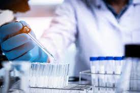
Doctoral student Jan-Samuel Puls. For this purpose, FtsZ is fluorescently labeled together with other proteins. The researchers then analyzed the effect on individual living bacterial cells over time, also using super-resolution microscopy. They built automated image analysis for microscope images, allowing them to quickly analyze all cells in a study sample. “Staphylococcus aureus is only about one micron, or one-thousandth of a millimeter. This makes microscopy particularly challenging,” says Dr.
Fabian Grein, junior research group leader at UKB’s Institute for Pharmaceutical Microbiology and scientist at the German Center for Infection Research (DZIF).
The Bonn research team discovered that the formation of peptidoglycan is the driving force for the entire cell division process. Previously, peptidoglycan synthesis was thought to be necessary only for a specific part of the process. The research team showed that inhibition of S. aureus cell wall assembly by glycopeptide antibiotics occurs rapidly and has a dramatic effect on cell division. In addition, they elucidated in detail the specific role of the essential penicillin-binding protein 2 (PBP2), which links cell wall components, in cell division. The beta-lactam antibiotic oxacillin prevented proper localization of this protein. “This means that PBP2 can’t get to where it’s needed. As a result, the cells can’t divide,” Grein said. “Importantly, this all happened immediately after the addition of antibiotics.
So, the first cellular effect, which has not been well studied so far, is crucial.” So given the alarming rate of antibiotic resistance worldwide growth, he hopes the findings will lead to a better understanding of how these drugs work at the cellular level, which could hold the key to developing new antibiotics.
Collected by CD BioGlyco, a biotechnology company that provides glycobiology-related products,analysis, custom synthesis, and design to advance glycobiology research. The company also providesPeptidoglycan-based Adjuvant Development, Peptidoglycan Structure Analysis, and Peptidoglycan Purification service for peptidoglycan research.